Topic of discussion: How the peripheral nerves fit in with the vertebral column.
Let’s start with the vertebral column and the structure of the vertebrae.
Vertebral Column Characteristics
-otherwise known as the spine
-made up of 26 irregular bones
-it is curved and flexible
-it starts at the skull and extends to the pelvis
-surrounds and protects the spinal cord
-ribs and muscles of the back and neck attach to it
-the sacrum and the coccyx are fused vertebrae, while the other 24 are separated by intervertebral discs.

www.academic.kellog.edu/herbrandsonc/bio201_McKinley
Divisions and Curvatures
The five major divisions are:
1. Cervical vertebrae, C1-C7 (seven vertebrae of the neck)
2. Thoracic vertebrae, T1-T12 (next 12 vertebrae)
3. Lumbar vertebrae, L1-L5 (5 vertebrae supporting the lower back)
4. Sacrum, S1-S5 (5 fused vertebrae)
5. Coccyx (terminus of the vertebral column, aka the tailbone)
The spine is curved like and “S”: the cervical and lumbar curvatures are concave and the thoracic and sacral curvatures are convex. This helps make the spine flexible.
Intervertebral Discs
-nucleus pulposus is the inner part of the disc. It makes the disc elastic and compressible.
-annulus fibrosus is the strong stuff surrounding of the nucleus pulposus. It is made up of collagen fibers and fibrocartilage.
-intervertebral discs act as shock absorbers and allow the spine to bend and flex.
 www.courses.vcu.edu
Structure of Vertebrae
-each has an anterior body (centrum) and a posterior vertebral arch.
-the body is disc shaped and it is the weight-bearing part.
-vertebral foramen is the opening surrounded by the body and arch.
-the spinal cord passes through the vertebral foramen and when stacked upon one another, it is called the vertebral canal.
-the vertebral arch is made up of 2 pedicles, and 2 laminae. The pedicles form the sides of the arch and the laminae complete the posterior side of the arch.
-there are 7 processes that project from the arch:
∙The spinous process and the transverse process are where muscles and ligaments attach.
∙A pair of superior articular processes and a pair of inferior articular processes form movable joints.
∙Intervertebral foramina are lateral openings between vertebrae, which is where the spinal nerves exit the vertebral column.

www.webschoolsolutions.com/patts/systems/vertebra
Moving on to the Peripheral Nerves……
2 Main parts of the Nervous System are:
-Central Nervous System (CNS) is the brain and spinal cord
-Peripheral Nervous System (PNS) is made up mostly of nerves that extend from the brain and the spinal cord.
Peripheral nerves carry messages to and from the CNS to all parts of the body.
2 Main parts of the PNS are:
Sensory (afferent) division:
-carries impulses from sensory receptors in the body to the CNS
-somatic sensory nerve fibers and visceral sensory nerve fibers
Motor (efferent) division:
-carries impulses from CNS to muscles and glands (effector organs)
-Motor division can be further divided into:
Somatic Nervous System – voluntary
-Carries impulses from CNS to skeletal muscles
Autonomic Nervous System – involuntary
-carries impulses from CNS to cardiac and smooth muscles, glands
-can be sympathetic (aka “fight or flight”) or parasympathetic (aka “resting and digesting”)
Neuroglia in PNS:
-Satellite cells: surround neuron cell bodies, function is unknown
-Schwann cells: insulate and form myelin sheaths around nerve fibers, vital to regeneration of nerve fibers www.steve.gb.com
Neurons (nerve cells):
-structural units of nervous system
-conduct nerve impulses
-neurons can live for a very longtime, given the right conditions
-amitotic, meaning they can’t divide, therefore they can’t be replaced
-require lots of oxygen and glucose
 www.home.ubalt.edu
Structure of a neuron:
Cell body (soma) - has a clear, round nucleus and most cell bodies are located in the CNS
Processes – arm-like extensions of the cell body. The PNS is mostly processes.
2 types of processes: dendrites and axons
Dendrites
-short, tapering, and diffusely branching = many per neuron
-bring incoming messages towards cell body
Axons
-one per neuron
-can be short or really, really long
-generates nerve impulses and transmits them away from cell body
 www.fusionanomaly.net/dendrite
Nerves:
-cordlike organ made up of a parallel bundle of peripheral axons wrapped in 3 layers of connective tissue: endoneurium, perineurium and epineurium.
-classified by the direction in which they transmit impulses
Sensory (afferent) nerves – carry impulses to CNS
Motor (efferent) nerves – carry impulses from CNS
Mixed nerves – have both sensory and motor fibers
-peripheral nerves are classified as cranial or spinal
-Spinal nerves carry impulses to and from the spinal cord and cranial nerves carry impulses to and from the brain.
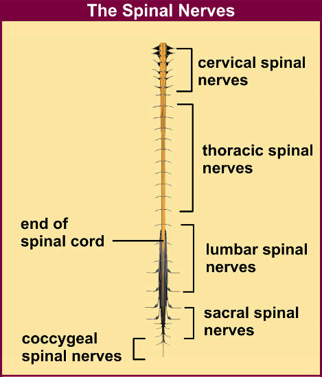
Spinal Nerves
-31 pairs with each made up of thousands of nerve fibers
-come from the spinal cord and go to all parts of the body except the head and some parts of the neck
-all are mixed nerves
-8 pairs of cervical nerves (C1-C8)
-12 pairs of thoracic nerves (T1-T12)
-5 pairs of lumbar nerves (L1-L5)
-5 pairs of sacral nerves (S1-S5)
-1 pair of coccygeal nerves (C0)
*there are 8 pairs of cervical nerves even though there are only 7 cervical vertebrae….the first 7 pairs exit superior to the vertebrae. Afterwards, all other spinal nerves exit inferior to the vertebrae.

www.spineuniverse.com www.becomehealthynow.com
Structure of the spinal nerve
-a dorsal root and a ventral root connect each spinal nerve to the spinal cord
-dorsal roots have sensory (afferent) fibers while ventral roots have motor (efferent) fibers
-spinal roots come out of the cord laterally and both roots unite to form the spinal nerve
-roots from the lumbar and sacral areas extend inferiorly through the cauda equina before they exit the vertebral column
-spinal nerves are short and after emerging from the intervertebral foramen, divide into dorsal ramus, ventral ramus and meningeal branch
-each ramus is made up of mixed nerves
Ventral rami branch and then form nerve plexuses that innervate the body.
There are cervical, brachial, lumbar and sacral plexuses.
Dorsal rami innervate the back by following the strip of muscle that is located in line with where it exits the vertebral column (so it follows the segmentation format!).
 www.training.seer.cancer.gov
No comments:
Post a Comment